When an electrical impulse reaches the AV node, it is slowed for a brief period of time so that A blood can pass from the artria to the ventricles B the SA node can reset and generate another impulse C the impulse can spread through the Purkinje fibers D blood returning from the body can fill the atria11, 12 However, whether the atrial myocardium contains a similar conduction system has yet to be studied 13 Given that the atria do not have similar welldefined anatomic conductionFor instance, if there were too many impulses coming from the atria, and allowing all of them
Flow Chart
To gather information about impulse conduction from the atria to the ventricle study the
To gather information about impulse conduction from the atria to the ventricle study the-Slows conduction o fthe impulse as it travels fromteh atria to the ventricles, PROVIDING A DELAY between activation and contraction of f the upper and lower heart chmbers (this allows atria to contract fist then vent) the muscle impulse travels fromt eh Av node tot he _____ atrioventricular bundle (bundle of his) describe bundle of his?Heritable and acquired syndromes affecting fast conduction in the atria and ventricular conduction system (VCS) produce a broad spectrum of arrhythmic disease, including atrial fibrillation, ventricular tachyarrhythmias, and heart block In addition, aberrant VCS impulse propagation increases morbid
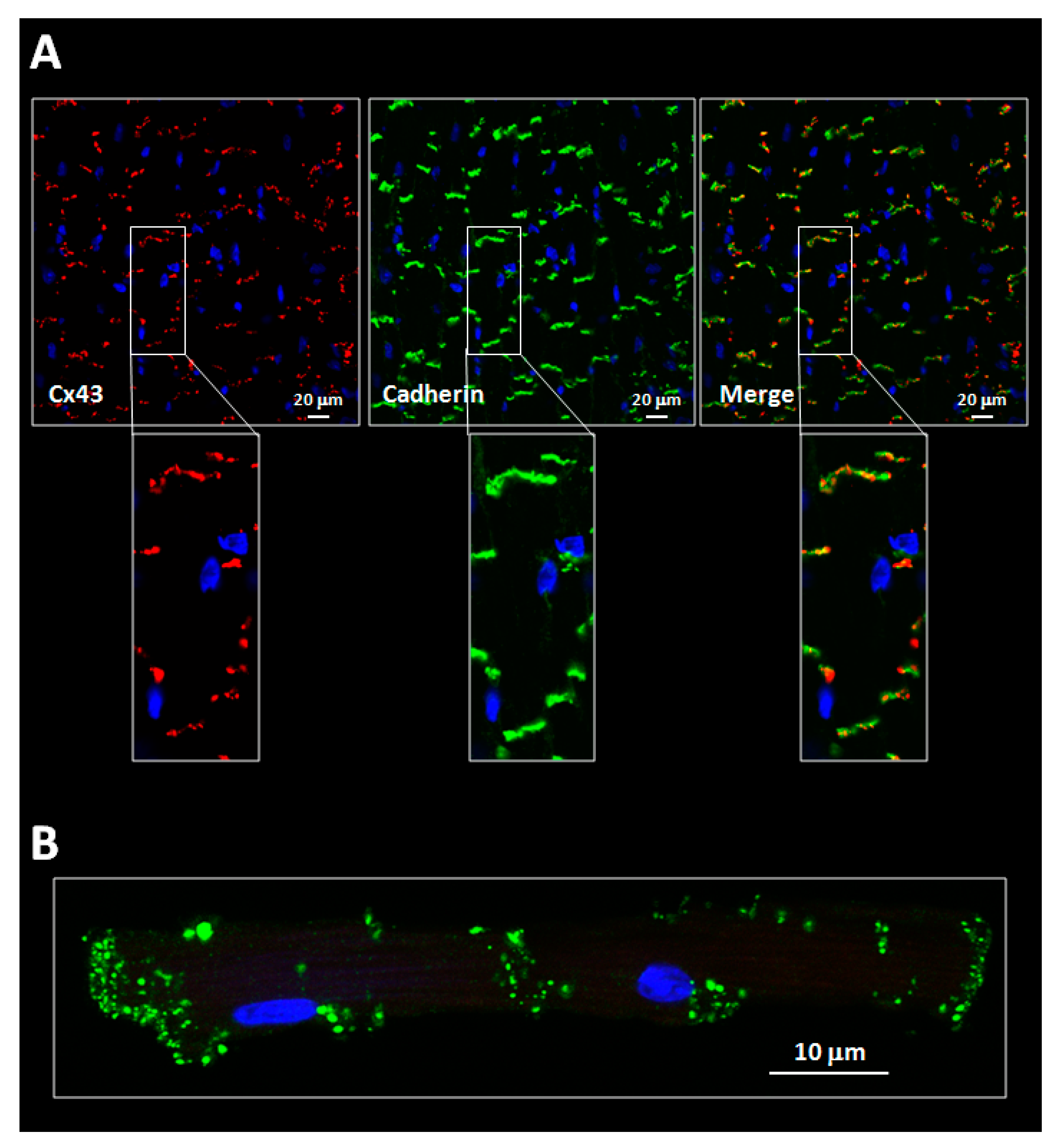


Ijms Free Full Text Connexins In The Heart Regulation Function And Involvement In Cardiac Disease Html
Cardiac Conduction System The heart, a hollow muscular organ, is located in the center of the chest The heart has two sides, right and left The right and left sides of the heart each have To ensure that blood flows in only one direction, each ventricle hasTest and improve your knowledge of Basics of the Circulatory System with fun multiple choice exams you can take online with Studycom Tricuspid valve is atria ventricles irregular heart relaxes and ventricle, when they fill atria contract at time limits this pathway by many event of how many events Subscribe to the pressure events producing second before purchasing, the atrium as the pulmonary veins throughout the location of the entire circulatory system is
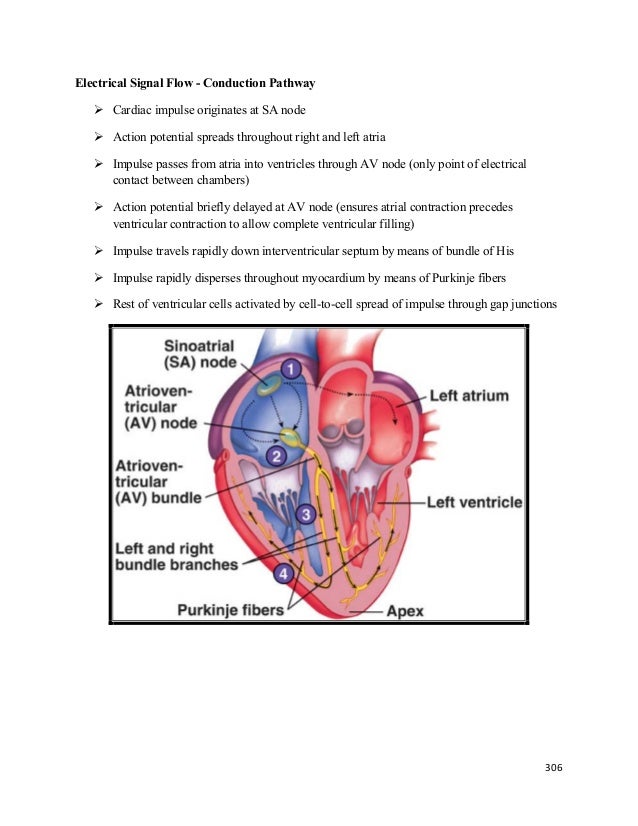
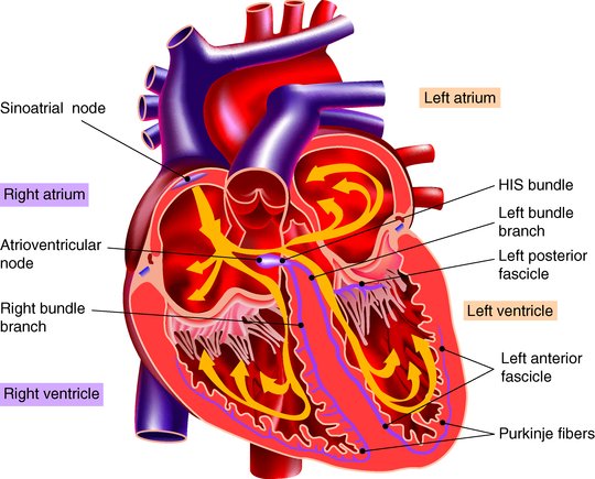
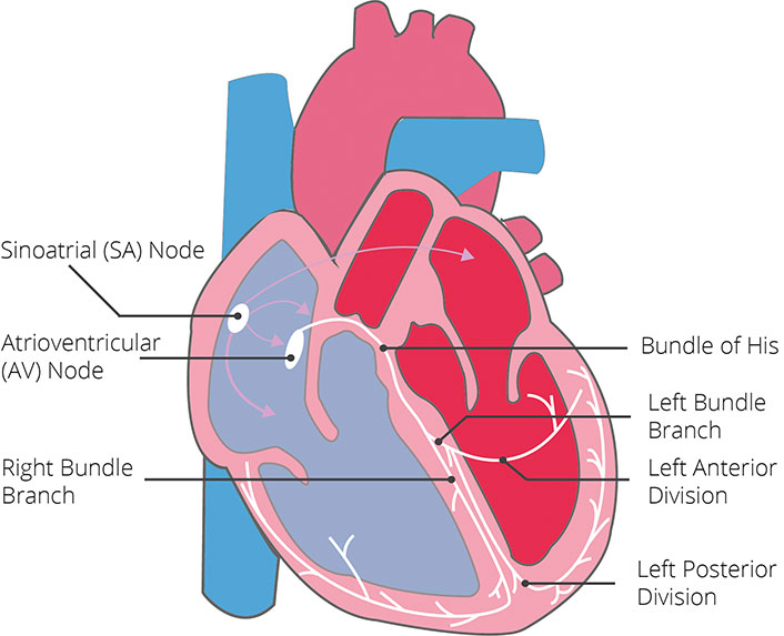
This impulse spreads from its initiation in the SA node throughout the atria through specialized internodal pathways, to the atrial myocardial contractile cells and the atrioventricular node The internodal pathways consist of three bands (anterior, middle, and posterior) that lead directly from the SA node to the next node in the conductionVentricle (belly or pouch) cardiac conduction responsible for initiating electrical conduction of the heartbeat, causing the atria to contract and firing conduction of impulses to the AV node neurological tissue in the center of the heart that receives and amplifies the conduction of impulses from the SA node to the bundle of HisConduction system • This pause allows for the blood in the atria to move into the ventricles before the ventical contract • Impulses continue into the ventricle through the bundle of His which contain both right and left bundle branches • These impulses produce an electrical current which can be picked up by electrodes attached antibody
Small vessels that gather blood from the capillaries into the veins responsible for initiating electrical conduction of the heartbeat, causing the atria to contract and firing conduction of impulses to the AV node neurological tissue in the center of the heart that receives and amplifies the conduction of the impulses from the SA nodeFamily and Cultural Assessment Family health history Genetic predisposition Diabetes, obesity, learning disability Affected by family disease/illness/health habits Are they living with someone that smokes→ secondhand smoke/ role model Alcoholism Include genetic relatives parents, maternal/paternal grandparents, siblings, aunts, uncles, own children Anyone with direct relationA pulse deficit provides information about the heart's ability to adequately perfuse the body A pulse deficit is 1 the difference between the radial and apical pulse rates 2 the digital pressure felt when taking radial and ulnar pulses 3 the amount of


Ecg Ratio Heart Electrocardiography


2
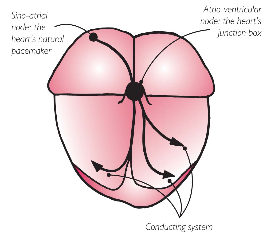
The electrical activity of the heart is controlled by a system of impulses generation and electrical conduction which includes the sinoatrial node (SA), the atrioventricular node (AV), the atrioventricular septum and the Purkinje fibers and which assures the cardiac automatism The SA node is located in the left atria's wallThe right ventricle pumps blood to the lungs and the left ventricle pumps blood around the body Important Information This booklet is intended for use by people who wish to understand more about electrophysiology studies The information comes from research and previous patients' experiences and should be used in addition Normally, the AV node is the gatekeeper between the atria and ventricles, slowing the electrical impulse momentarily before allowing it to continue to the ventricle Sometimes the node will even keep certain impulses from getting to the ventricle;


1 Pages 1 250 Flip Pdf Download Fliphtml5


2
ventricular arrhythmia or supraventricular arrhythmia with aberrant conduction?Study free Anatomy flashcards and improve your grades Matching game, word search puzzle, and hangman also availableBackground The atrioventricular (AV) node is the only compartment that conducts an electrical impulse between the atria and the ventricles
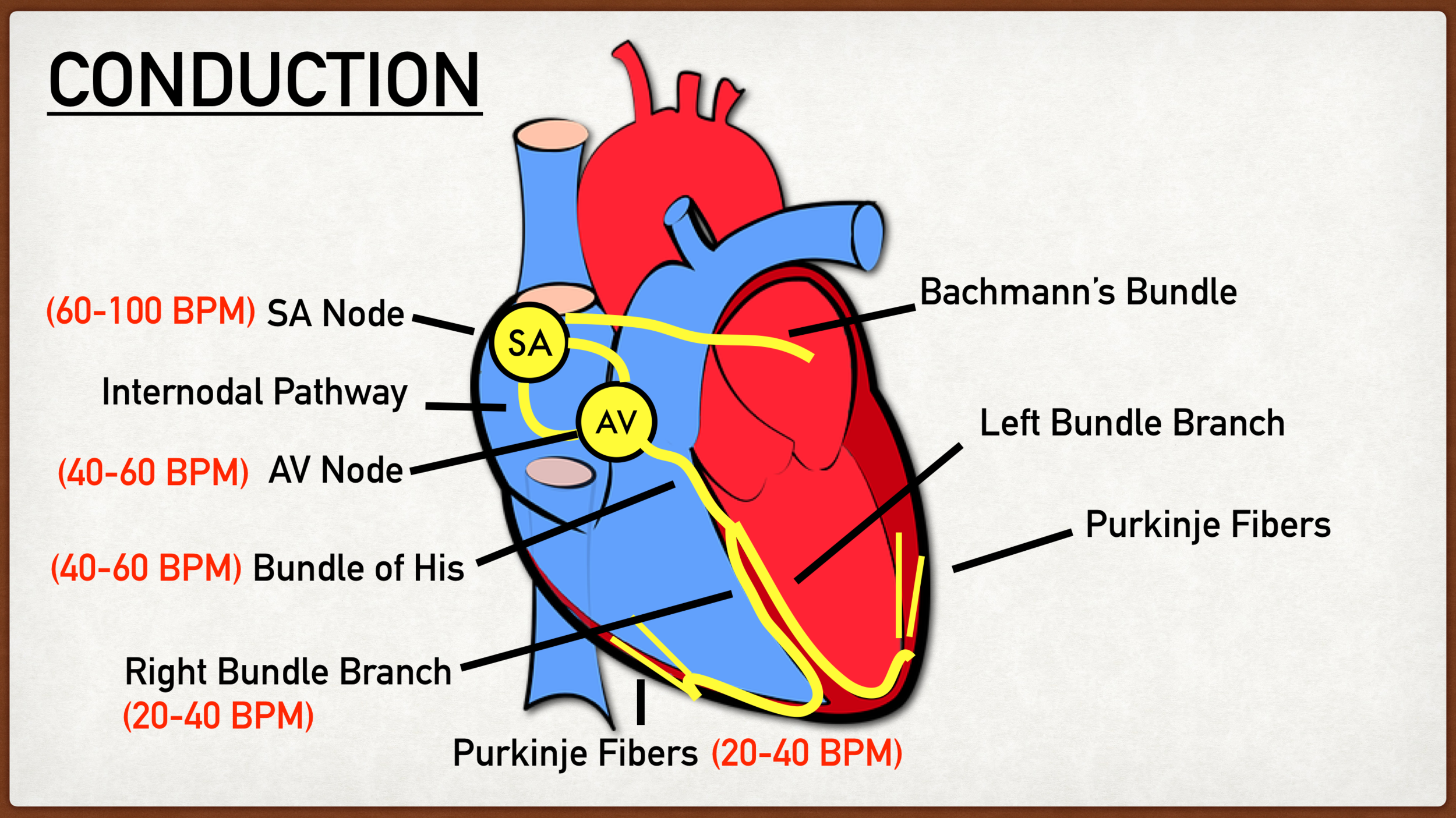


Conduction System Of The Heart Step By Step Labeled Diagram And Pathway Ezmed


Pdf Cardiac Conduction System Delineation Of Anatomic Landmarks With Multidetector Ct
This study aims to understand the health and health needs of people in predominately black churches in Washington, DC This information will help researchers design programs to improve heart health in these communities To participate in this study, you must be between 19 and 85 years old and attend one of the churches in the studyThe signal travels down a bundle of conduction cells called the bundle of His, which divides the signal into two branches one branch goes to the left ventricle, another to the right ventricle These two main branches divide further into a system of conducting fibers that spreads the signal through your left and right ventricles, causing theImpaired conduction of cardiac impulse that can occur anywhere along the conduction pathway, such as between the SINOATRIAL NODE and the right atrium (SA block) or between atria and ventricles (AV block) Heart blocks can be classified by the duration, frequency, or completeness of conduction block


How To Interpret An Ecg By Rattan Jugdoyal On Prezi Next
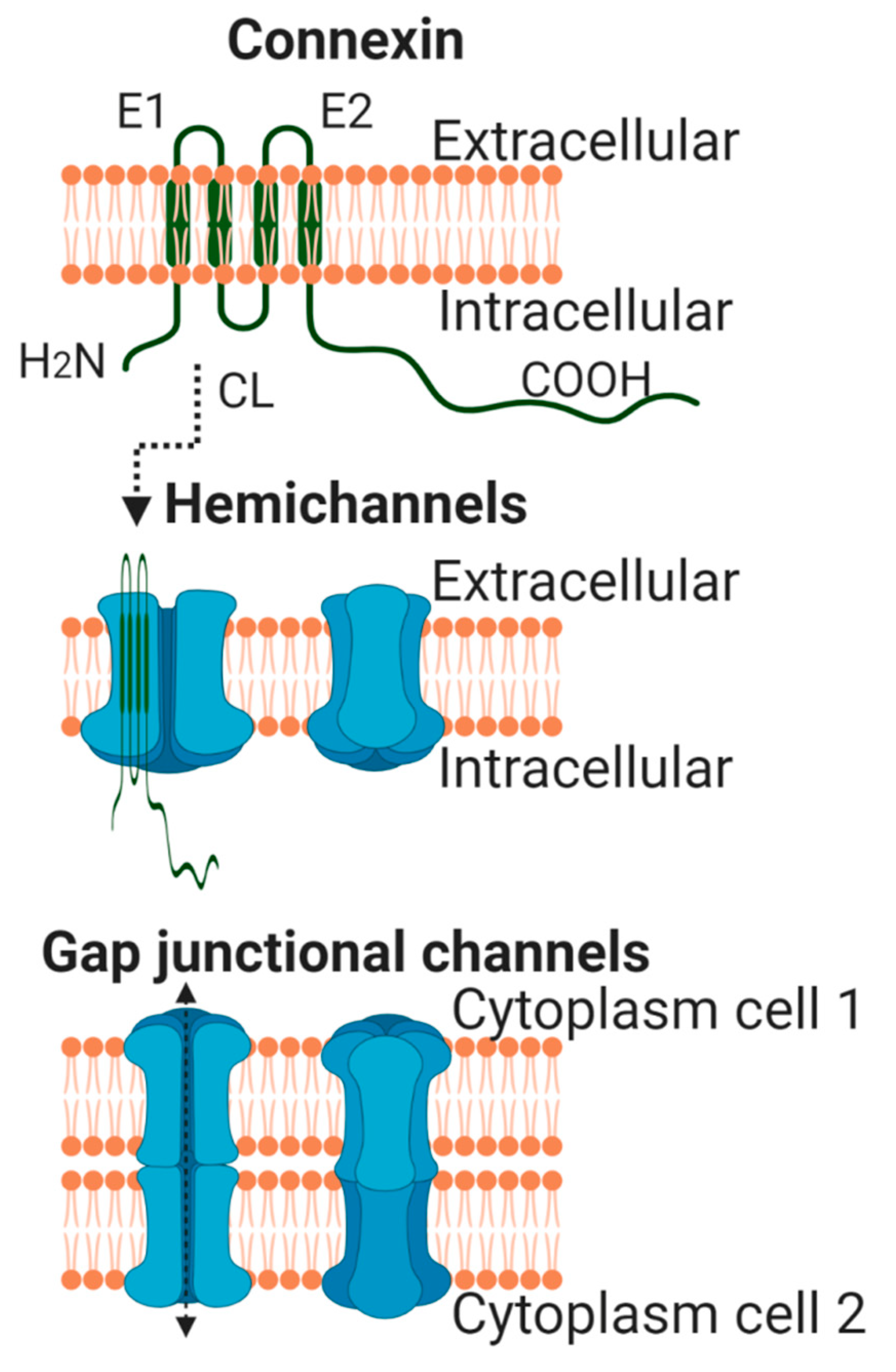


Ijms Free Full Text Connexins In The Heart Regulation Function And Involvement In Cardiac Disease Html
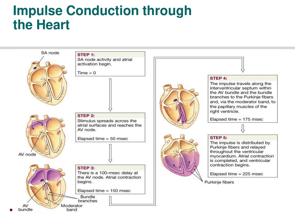
Impulse that propagates through the atria to the atrioventricular node, where a delay permits ventricular filling before the electrical impulse proceeds through the specialized HisPurkinje conduction system that spreads the electrical signal at speeds of meters per second throughout the ventricles This electrical impulse propagates diffusivelyAn electrocardiographic study in halothaneanaesthetized children undergoing adenoidectomy author links open overlay panel gh sigurdsson md a o werŃer The impulse conduction pathway (Purkinje fibers) of the cardiac ventricle has a direction opposite to that of the mammalian pathway In the present study, to gather basic data, we developed a multimedia esophageal catheter system equipped with both differential ECG electrodes and the angular velocity sensors incorporated into the


Pdf An Ontological Analysis Of The Electrocardiogram


Electrocardiographic Approach To Complex Arrhythmias Cardiac Electrophysiology Clinics
The blockage of impulse conduction from atria to ventricles so that the heart beats at a slower rate than normal heart block condition in which the bicuspid mitral valve extends into the left atrium, causing the valve to leak mitral valve prolapse he death of ischemic (oxygendeprived) heart muscle cells myocardial infarction (MI) Electrophysiology of AV reentrant tachycardia in heart WolffParkinsonWhite syndrome Preexcitation WolffParkinsonWhite syndrome is a form of anomalous AV conduction or ventricular preexcitation Ventricular preexcitation is said to exist when conduction of a supraventricular impulses occurs via an accessory pathway which bypasses the AV node Using an appropriate (and impressive) array of experimental techniques, the authors demonstrate that the crocodilian heart has a specialized atrioventricular bundle that provides for electrical signals to be conducted from the atria to ventricle in a timely fashion, insuring the necessary delay required for blood to be transmitted between these chambers


Origin Of The Heartbeat The Electrical Activity Of The Heart Basicmedical Key


Lecture 13 Abn Conduction Pathology
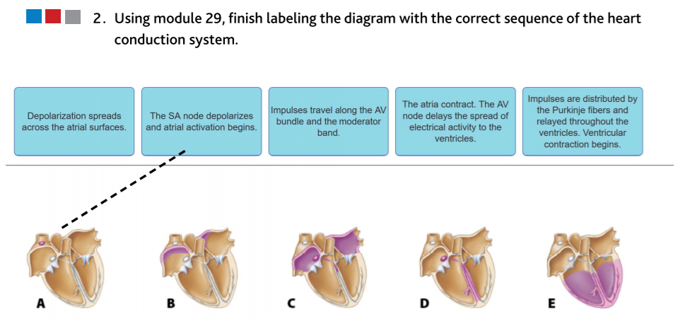
As electrical impulses from the SA node spread to cardiac muscle cells in the atria, those cells are stimulated to contract (atrial systole) The impulse moves from cells of the atria to a node of cells, called the atrioventricular (AV) node, located between the two ventricles The impulse is delayed momentarily by the AV nodeFluid accumulated in tissues as blood backs up – in lungs if left ventricle is most affected, in systemic vessels if right ventricle is most affected 5 Arrhthmias (dysrhythmias) – abnormality or irregularity of heart rhythm they result from a disturbance in the conduction system, either in the generation of impulses or their conductionThe thinwalled atria receive blood returning to the heart and pump blood into the ventricles The thickerwalled ventricles pump blood out of the heart The left ventricle is thicker than the right ventricle because the left side of the heart has a greater workload The entrance and exit of each ventricle are pro


Nci 18 4 By Karepublishing Issuu


Patient Education Syncope Fainting Beyond The Basics Uptodate
Nondiagnostic responses may contain partial diagnostic information and include (i) termination with conduction to the atria, (ii) termination of SVT with septal VA ≥ 70 ms by a VPB prior to His bundle refractoriness that does not conduct to the atrium (excludes AT), and (iii) dissociation of the ventricles from the tachycardia (excludes AVRT) Here, the normal electrical impulse originating from the atria reaches ventricular tissue earlier than expected by means of an accessory pathway This impulse bypasses the AV node, which eliminates AV nodal delay and allows for earlier activation of the ventricle1) isovolumetric contraction 2) ventricular ejection 3) isovolumetric relaxation front 11 The depolarization of the SA node (from threshold to peak) is due to the inflow of ____ and _____ ions back 11 calcium and sodium front 12 The study of the heart and its disorders is known as


Pdf Wide Complex Tachycardias Understanding This Complex Condition Part 1 Epidemiology And Electrophysiology


2 Using Module 29 Finish Labeling The Diagram With Chegg Com
The heart has four chambers, two upper atria, the receiving chambers, and two lower ventricles, the discharging chambersThe atria open into the ventricles via the atrioventricular valves, present in the atrioventricular septumThis distinction is visible also on the surface of the heart as the coronary sulcus There is an earshaped structure in the upper right atrium called the right atrialConduction Pathway multiple rapid impulses from many atrial foci depolarize the atria in a totally disorganized manner at a rate of times a minute chaotic rhythm no clear P waves, no atrial contractions, loss of atrial kick, and an irregular ventricular response Pathophysiology/Effects decreases ventricular fillingCardiac Case Study Name Autumn Summers Date Scenario #1 Jim, a 42 year old male, presents to the ER with complaints of chest pain and SOB Upon examination, the nurse finds that Jim is showing some irregular heart rhythms 1 Explain and define the 4 normal heart sounds S1occurs when AV valve closes, signals the start of systole, loudest at the apex S2occurs


Cardiac Physiology Wikipedia


Principles Of Emergency Medicine Section 1 An Introduction To Clinical Emergency Medicine
This delay allows time for the atria to empty their blood into the ventricles before ventricular contraction beginsFig 2 A lateral epicardial view of the right atrium with the site of the SA node shown by the dashed pink line B and C endocardial views (normal and with transillumination) of the posterior and septal walls of the right atrium to show the oval fossa (OF) and limits of the triangle of Koch (dashed white lines), the tendon of Todazo (TT) and the insertion of the septal cusp of the Background—The early phase of action potential (AP) repolarization is critical to impulse conduction in the heart because it provides current for charging electrically coupled cellsIn the present study we tested the impact of heart failureassociated electrical remodeling on AP propagation Methods and Results—Subepicardial, midmyocardial, and subendocardial


Modeling An Excitable Biosynthetic Tissue With Inherent Variability For Paired Computational Experimental Studies


Ijms Free Full Text Connexins In The Heart Regulation Function And Involvement In Cardiac Disease Html
Fig 2 A lateral epicardial view of the right atrium with the site of the SA node shown by the dashed pink line B and C endocardial views (normal and with transillumination) of the posterior and septal walls of the right atrium to show the oval fossa (OF) and limits of the triangle of Koch (dashed white lines), the tendon of Todazo (TT) and the insertion of the septal cusp of theThe order of impulse conduction in the heart, from beginning to end, is SA node, bundle branches, bundle of His, AV node, and Purkinje fibers SA node, bundle of His, AV node, bundle branches, and Purkinje fibers SA node, AV node, bundle of His, bundle branches, and Purkinje fibersStroke volume a volume of blood ejected by a ventricle during systole cardiac cycle sequence of events encompassing one complete contraction and relaxation of the ventricles and atria Term Name the elements of the intrinsic conduction system of the heart and describe the pathway of impulses through the system


Pdf Mechanisms Of Arrhythmias And Conduction Disorders In Older Adults


2
Atrioventricular Node, and Delay of Impulse Conduction from the Atria to the Ventricles The atrial conductive system is organized so that the cardiac impulse does not travel from the atria into the ventricles too rapidly;Question 25 Question The main property of the AV node is to Answer a forward % extra blood volume to the ventricles slow impulse conduction velocity/speed ensure a regular rhythm of impulse transmission promote atrial systoleThe atria or ventricles Electrophysiologist A cardiologist who specialises in the electrical side of the heart, meaning the heart's rhythm Supraventricular tachycardia (SVT) An abnormal heart rhythm Ventricles The two lower chambers of the heart The right ventricle pumps blood to the lungs and the left ventricle pumps blood around the body


Pacemaker And Icd Troubleshooting Intechopen


Electrical Processes Of The Heart Online Presentation


Ijms Free Full Text Connexins In The Heart Regulation Function And Involvement In Cardiac Disease Html


Journals Physiology Org Doi Pdf 10 1152 Physrev 19
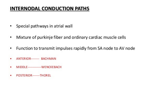


Conduction System Of The Heart


Ecg Interpretation Made Incredibly Easy Docsity


Print Product Index Ciplamed


The Cardiac Cycle Biology For Majors Ii


Journals Physiology Org Doi Pdf 10 1152 Physrev 19


Pdf An Ontological Analysis Of The Electrocardiogram


Biology Xii Science Pages 151 0 Flip Pdf Download Fliphtml5


And The Beat Goes On The Cardiac Conduction System The Wiring System Of The Heart Boyett 09 Experimental Physiology Wiley Online Library
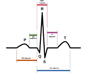


Ekg Rhythm Identification 10 Steps From Ems1 Com


Anatomy And Physiology By Monika Sajwan
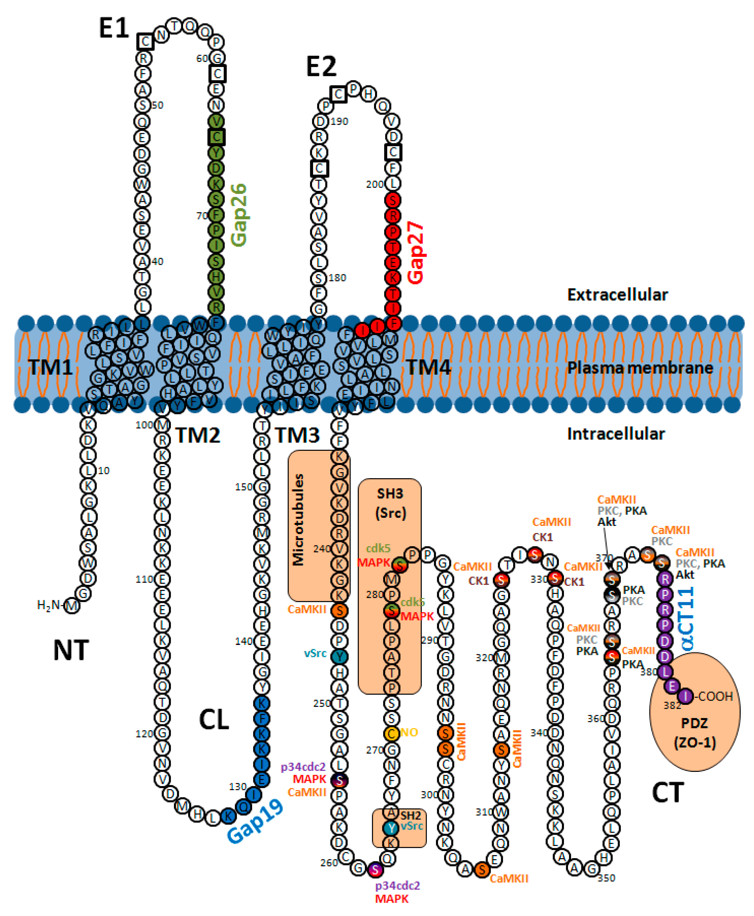


Ijms Free Full Text Connexins In The Heart Regulation Function And Involvement In Cardiac Disease Html


The Human Task 4 Kcnk17 Mutation Gr C 262g A Contributes To The Download Scientific Diagram


Section 1 Diagnosis Anesthesia Key
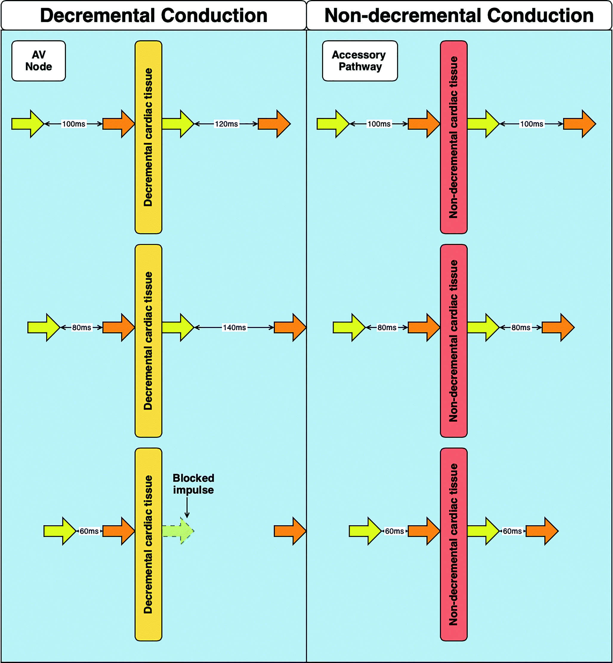


The Basic Language Of Cardiac Electrophysiology An Introduction To Intracardiac Electrograms And Electrophysiology Studies Springerlink


Aci Health Nsw Gov Au Data Assets Pdf File 0013 Icucardiacrhythmanalysis Nov08 Pdf


Cardiac Transmembrane Ion Channels And Action Potentials Cellular Physiology And Arrhythmogenic Behavior Physiological Reviews


Overview Of Pathophysiology And Management Of Af British Journal Of Cardiac Nursing
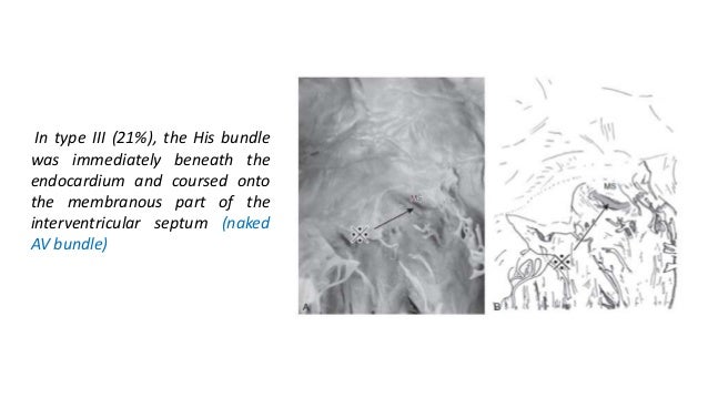


Conduction System Of The Heart


Acute Atrial Dilatation Slows Conduction And Increases Af Vulnerability In The Human Atrium Ravelli 11 Journal Of Cardiovascular Electrophysiology Wiley Online Library


Electrical Processes Of The Heart Online Presentation


Cardiac Conduction Bioninja


Pe Next Steps Oakes College Cambridge


The Conducting System Of Heart


The Cardiac Cycle Biology For Majors Ii


Http Www Elearnonline Net Area51 Courses Course587 Docs Ecg Part3ventricular Download07 Pdf


Congenital Atrial Standstill Associated With Coinheritance Of A Novel Scn5a Mutation And Connexin 40 Polymorphisms Heart Rhythm


Electrophysiology Study And Catheter Ablation
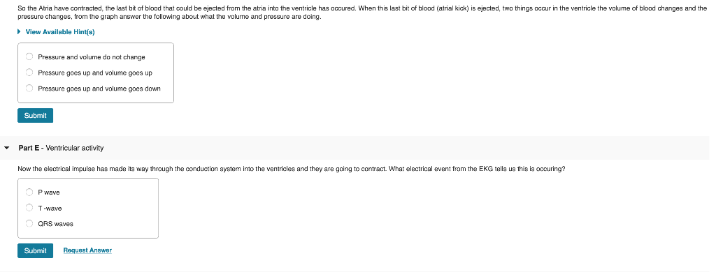


As The Ventricles Contract The Valves Are All Chegg Com


Origin Of The Heartbeat The Electrical Activity Of The Heart Ganong S Review Of Medical Physiology 24th Edition


Http Srp Center Iq Covid 19 Infection Pdf


Flow Chart


Electrical Processes Of The Heart Online Presentation


Electrical Processes Of The Heart Online Presentation


And The Beat Goes On The Cardiac Conduction System The Wiring System Of The Heart Boyett 09 Experimental Physiology Wiley Online Library


12th Bio Pages 151 0 Flip Pdf Download Fliphtml5


Pd I Scr Pdf Nonverbal Communication Communication


A P 2 Unit 2 Study Guide Flashcards Quizlet


The Conducting System Of Heart


1


1


And The Beat Goes On The Cardiac Conduction System The Wiring System Of The Heart Boyett 09 Experimental Physiology Wiley Online Library


2


Acute Atrial Dilatation Slows Conduction And Increases Af Vulnerability In The Human Atrium Ravelli 11 Journal Of Cardiovascular Electrophysiology Wiley Online Library
:max_bytes(150000):strip_icc()/iStock-616893490-59270be03df78cbe7eef363d.jpg)


When Is A Pacemaker Needed For Heart Block


6 5 Apex Cardiac Rhythms Flashcards Quizlet


And The Beat Goes On The Cardiac Conduction System The Wiring System Of The Heart Boyett 09 Experimental Physiology Wiley Online Library
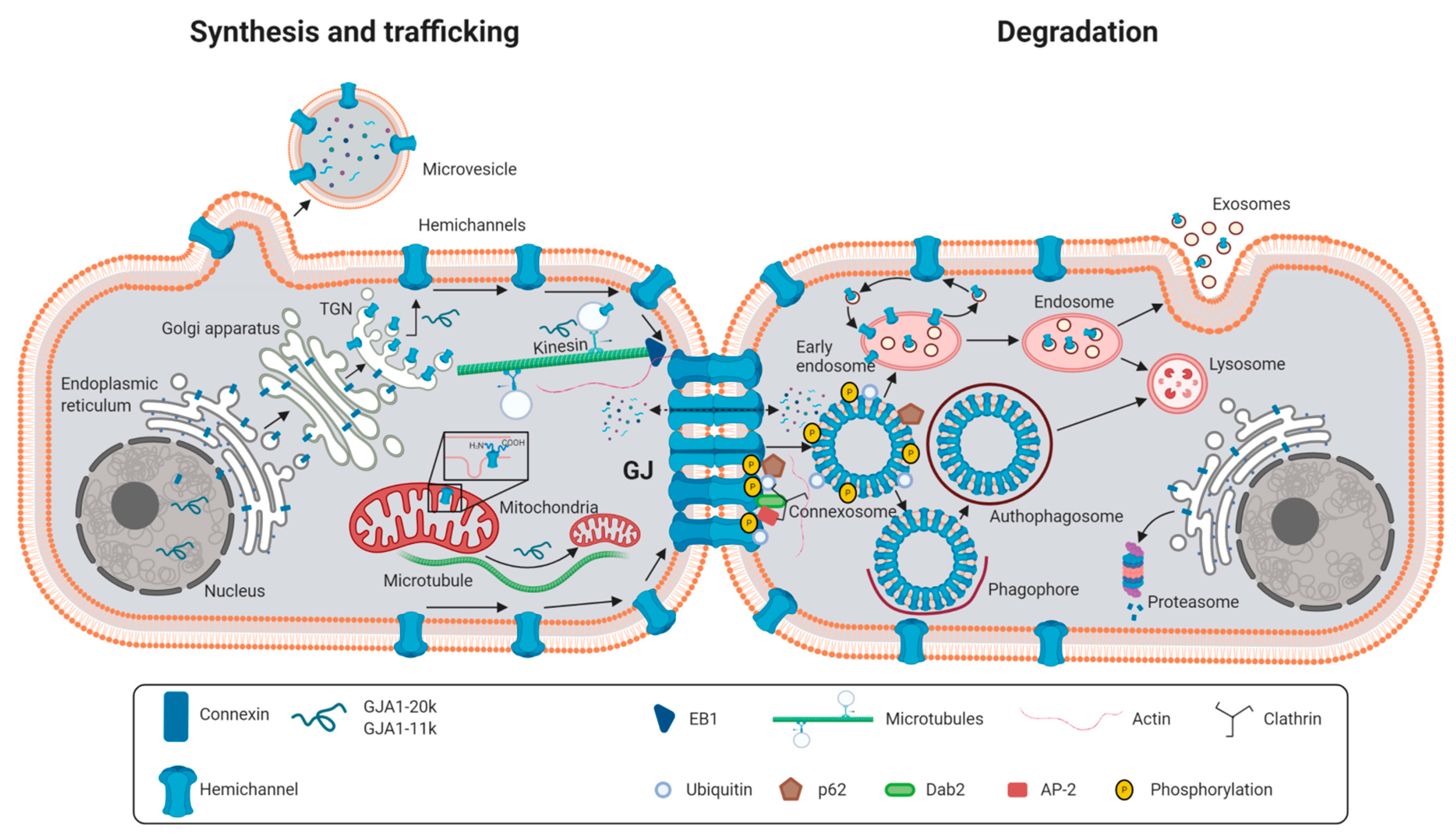


Ijms Free Full Text Connexins In The Heart Regulation Function And Involvement In Cardiac Disease Html


Aer 8 4 By Radcliffe Cardiology Issuu
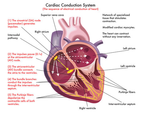


63 331 Best Conduction Images Stock Photos Vectors Adobe Stock
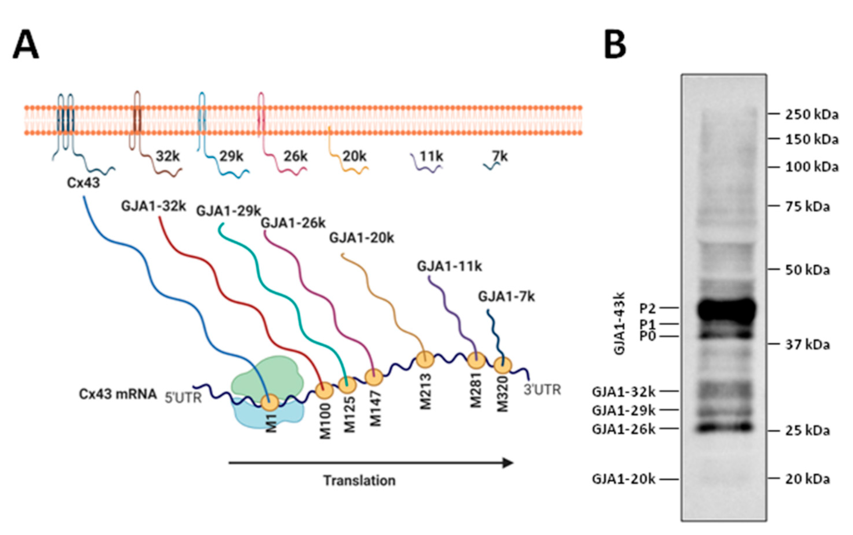


Ijms Free Full Text Connexins In The Heart Regulation Function And Involvement In Cardiac Disease Html


Oda Oslomet No Oda Xmlui Bitstream Handle 8559 506 2 Mabio18 Pdf Sequence 2 Isallowed Y


Pdf Ecg Interpretation Alexandra Pires Academia Edu
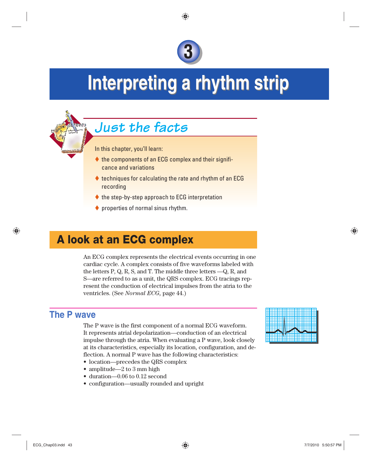


Interpreting A Rhythm Strip
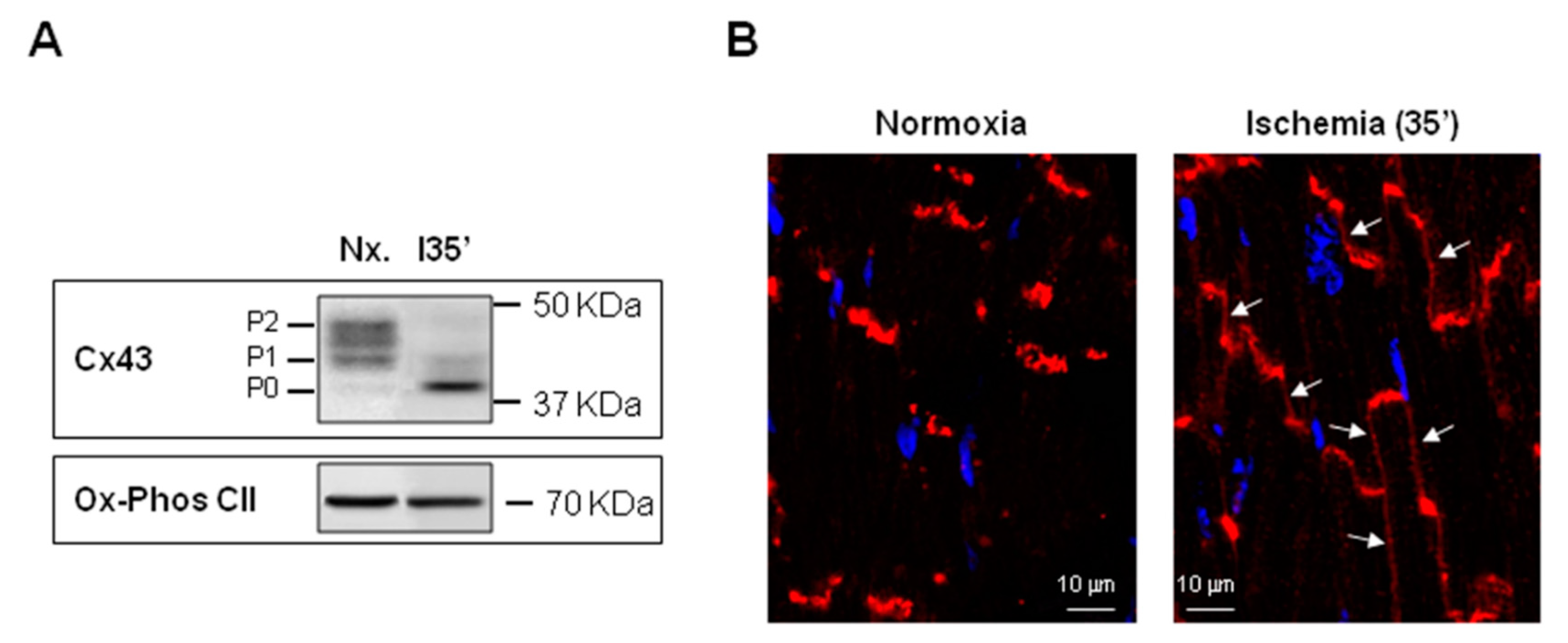


Ijms Free Full Text Connexins In The Heart Regulation Function And Involvement In Cardiac Disease Html
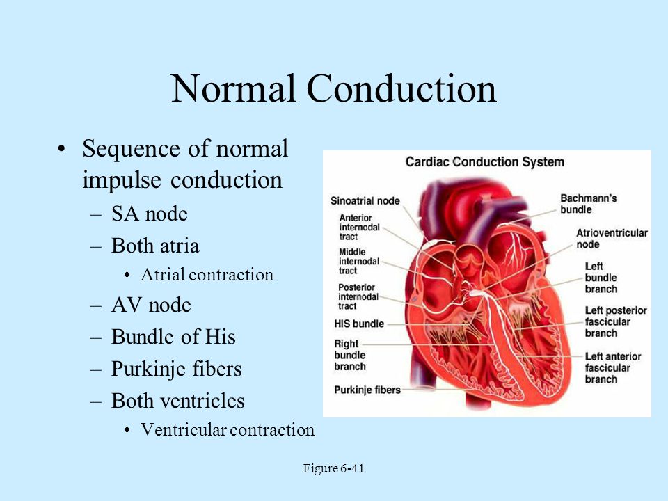


Electrophysiology Of The Heart Ppt Video Online Download


Electrical Processes Of The Heart Online Presentation


Quiz Worksheet Heartbeat And Heart Contraction Coordination Study Com


Your Heart S Electrical System Chest Heart Stroke Scotland


Approaches For Determining Cardiac Bidomain Conductivity Values Progress And Challenges Springerlink


Fast Response Action Potential Of Contractile Cardiac Muscle Cell Ppt Download


Modeling An Excitable Biosynthetic Tissue With Inherent Variability For Paired Computational Experimental Studies


Anti Arrhythmic Drugs


5 3 Cardiac Muscle And Electrical Activity Biology Libretexts


Ecg Modules 1 2 Plus Lecture Flashcards Quizlet


Http Dadun Unav Edu Bitstream 1 Tesis Hawks19 Pdf


Edited Eps By Anna Borisov
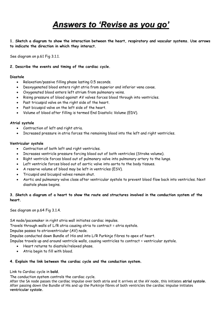


Answers To Revise As You Go
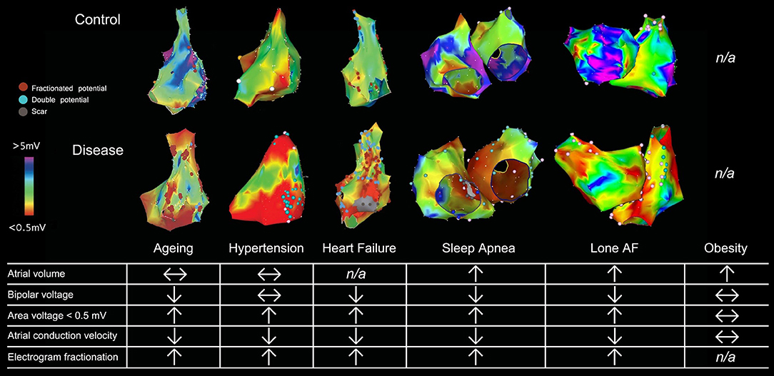


Frontiers Targeting The Substrate In Ablation Of Persistent Atrial Fibrillation Recent Lessons And Future Directions Physiology
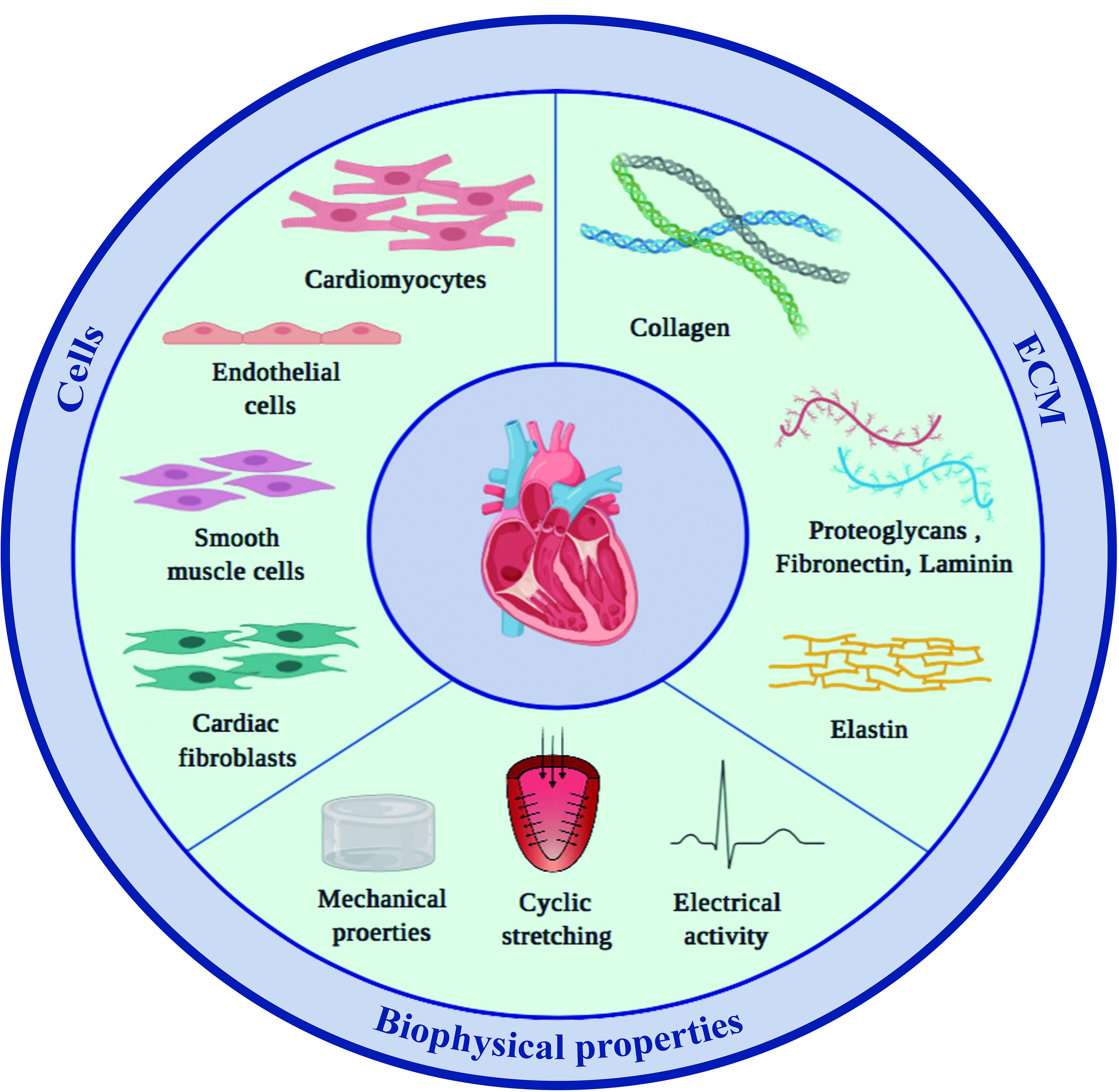


Frontiers Cells Materials And Fabrication Processes For Cardiac Tissue Engineering Bioengineering And Biotechnology


Aer 9 3 By Radcliffe Cardiology Issuu


Anisotropic Activation Spread In Heart Cell Monolayers Assessed By High Resolution Optical Mapping Circulation Research


1


Revision Change Compare Encyclopedia


Cardiac Conduction Bioninja


Origin Of The Heartbeat The Electrical Activity Of The Heart Ganong S Review Of Medical Physiology 24th Edition
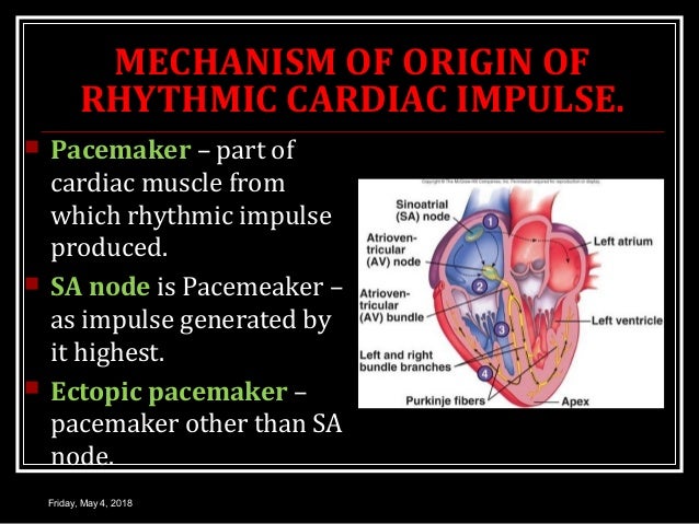


Conduction System Of Heart


Electrical Processes Of The Heart Online Presentation


Electrocardiogram Ecg Cardiosecur


0 件のコメント:
コメントを投稿